Associated Toxicities: Assessment and Management Related to CAR T-Cell Therapy
Background: The impressive disease response observed with chimeric antigen receptor (CAR) T-cell therapy is accompanied by the potential for unique and severe toxicities. Cytokine release syndrome (CRS) and neurologic toxicities have emerged as prominent toxicities associated with this treatment modality.
Objectives: This article presents an overview of pathophysiology, assessment, and evidence-based management of CAR T-cell therapy–associated toxicities, with particular attention paid to CRS and neurologic toxicity management. Implications for nursing practice are included for prominent toxicities to guide clinical practice.
Methods: An overview of recent guidelines and evidence for CAR T-cell therapy toxicity assessment and management is provided.
Findings: Evidence-based approaches to CAR T-cell therapy toxicities continue to evolve. As organizational and institutional guidelines emerge, nurses must be aware of anticipated toxicities and interventions used in clinical practice to provide timely and effective care.
Jump to a section
Chimeric antigen receptor (CAR) T-cell therapy targeting CD19 has been associated with dramatic treatment responses in B-cell acute lymphoblastic leukemia (ALL) and large B-cell lymphomas. However, management of treatment-related toxicities is a significant concern (Abramson et al., 2018; Maude et al., 2018; Neelapu et al., 2017; Schuster et al., 2017). Black-box warnings for cytokine release syndrome (CRS) and neurologic toxicities exist for U.S. Food and Drug Administration (FDA)–approved CAR T-cell agents (i.e., axicabtagene ciloleucel [Yescarta®] and tisagenlecleucel [Kymriah®]) because of their potential for life-threatening or fatal side effects. Both products are available only through Risk Evaluation and Mitigation Strategy (REMS) programs (Kite Pharma, 2018; Novartis Pharmaceuticals, 2018b). REMS programs require authorized centers to comply with specific guidelines to mitigate the risks of treatment. Hospitalization and intensive care unit admission for treatment of CRS and neurologic toxicity symptoms may be needed. Evidence for best management strategies continues to evolve; however, specific interventions may vary based on institutional practice. Toxicity consensus guidelines have been published and are being translated into clinical practice (Lee et al., 2018; Mahmoudjafari et al., 2019; Neelapu et al., 2018; Teachey, Bishop, Maloney, & Grupp, 2018).
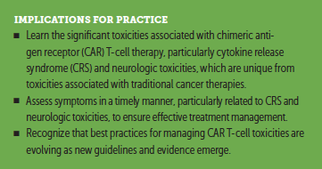
In addition to CRS and neurologic toxicity, other side effects related to CD19-directed CAR T cells include cytopenias, infections, hypogammaglobulinemia, and tumor lysis syndrome (TLS) (Brudno & Kochenderfer, 2016). Education for the interprofessional CAR T-cell team, as well as for patients and caregivers, is essential to ensure that toxicities are appropriately monitored and managed. Nurses are pivotal in assessing, identifying, and managing toxicities to promote best patient outcomes.
Cytokine Release Syndrome
CRS is the most common toxicity associated with CAR T-cell therapy (Brudno & Kochenderfer, 2016). CRS is a systemic inflammatory response involving elevated cytokines that occurs with immune system activation (Lee et al., 2014; Wang & Han, 2018). As CAR T cells are activated and proliferate, cytokines are released, stimulating other cells in the immune system, such as macrophages and endothelial cells (Shimabukuro-Vornhagen et al., 2018; Wang & Han, 2018). Elevated cytokines include tumor necrosis factor alpha, interleukin-2, interleukin-6 (IL-6), interferon gamma, granulocyte macrophage–colony-stimulating factor (GM-CSF), and interleukin-8 (Brudno & Kochenderfer, 2018; Lee et al., 2014). IL-6 is thought to be associated with peak CRS toxicity (Lee et al., 2014).
Fever is the hallmark sign of CRS, and symptoms can appear similar to infection. Fevers may reach as high as 40ºC–41ºC. Symptoms such as tachycardia, chills, myalgias, arthralgias, malaise, and fatigue may also present (Brudno & Kochenderfer, 2016; Lee et al., 2014). More severe CRS symptoms include hypotension, dyspnea, hypoxia, respiratory distress, coagulopathies, and organ toxicities, such as cardiac, renal, and liver dysfunction (Brudno & Kochenderfer, 2016; Hay et al., 2017). Severe CRS may prompt symptoms consistent with macrophage activation syndrome and hemophagocytic lymphohistiocytosis (Neelapu et al., 2018). Life-threatening complications can include cardiac dysfunction (e.g., arrhythmias, cardiomyopathy), adult respiratory distress syndrome, renal or hepatic failure, and disseminated intravascular coagulation (Brudno & Kochenderfer, 2016; Lee et al., 2014).
Median onset of CRS for FDA-approved CAR T-cell agents is two to three days following infusion, with symptoms usually presenting within the first one to two weeks following infusion (Kite Pharma, 2017; Maude et al., 2018; Neelapu et al., 2017; Novartis Pharmaceuticals, 2018a). The development of CRS may be influenced by disease burden at the time of treatment, CAR T-cell type, and cell dose (Brudno & Kochenderfer, 2018; Shimabukuro-Vornhagen et al., 2018).
Multiple grading scales have been developed by consensus groups or institutions to guide CRS assessment and management, and these have been described extensively elsewhere (Brudno & Kochenderfer, 2018; Kite Pharma, 2017; Lee et al., 2014, 2018; Mahadeo et al., 2019; Neelapu et al., 2018; Novartis Pharmaceuticals, 2017a; Park et al., 2018; Porter, Frey, Wood, Weng, & Grupp, 2018; Santomasso et al., 2018; University of Texas MD Anderson Cancer Center, 2017). The Lee et al. (2014) scale has been used widely for grading CRS in clinical trials, including the clinical trials for axicabtagene ciloleucel (Neelapu et al., 2017). The University of Pennyslvania developed a separate grading scale for CRS (Penn scale), which was used in clinical trials for tisagenlecleucel (Porter et al., 2018). Differences exist between the Lee et al. (2014) scale and the Penn scale, particularly regarding the use of vasopressors, making comparison of toxicities across trials challenging. For example, a patient with CRS-associated hypotension responsive to low-dose vasopressors could be classified as grade 2 CRS on the Lee et al. (2014) scale and grade 3 on the Penn scale. Consensus guidelines, such as those published by the multi-institution and multidisciplinary CARTOX (CAR T-cell therapy–associated toxicity) Working Group and the American Society for Blood and Marrow Transplantation (ASBMT), have been developed in attempts to further standardize grading and toxicity management in clinical practice (Lee et al., 2018; Mahadeo et al., 2019; Neelapu et al., 2018). In general, CRS is graded on a scale of 1–4, with a grade of 3 or higher indicating severe symptoms (Lee et al., 2014, 2018; Mahadeo et al., 2019; National Cancer Institute Cancer Therapy Evaluation Program, 2018; Neelapu et al., 2018; Porter et al., 2018). The grade is determined by a combination of clinical symptoms, including fever, hypotension, hypoxia, and organ dysfunction.
Management Strategies
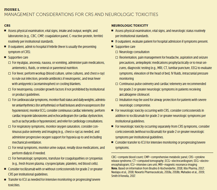
Management of grade 1 (mild) CRS is supportive and consists of managing flu-like symptoms, myalgias, headache, nausea, and fatigue (Lee et al., 2014; Neelapu et al., 2018). Grade 2 or higher CRS involves management of hypotension, hypoxia, and organ dysfunction with fluids, vasopressors, oxygen, and other supportive interventions (Brudno & Kochenderfer, 2018; Mahadeo et al., 2019; Neelapu et al., 2018). An overview of common management strategies, adapted from the drug prescribing information and other published guidelines, is presented in Figure 1. 
Tocilizumab (Actemra®) is a humanized monoclonal antibody that blocks binding to IL-6 receptors and is approved by the FDA for the treatment of CRS (Genentech, 2018). Tocilizumab administration can lead to rapid improvement or resolution of CRS symptoms in many cases (Brudno & Kochenderfer, 2016). Tocilizumab may be ordered for grade 2 CRS symptoms, such as hypoxia or hypotension, that are not responding to supportive care interventions, such as fluids and oxygen (Kite Pharma, 2017; Lee et al., 2014; Neelapu et al., 2018; Novartis Pharmaceuticals, 2018a; Porter et al., 2018). For severe (grade 3 or greater) CRS symptoms, corticosteroids are usually prescribed in addition to tocilizumab (Kite Pharma, 2017; Lee et al., 2014; Neelapu et al., 2018; Novartis Pharmaceuticals, 2018a; Porter et al., 2018). Tocilizumab is administered intravenously over 1 hour at a dose of 8 mg/kg for patients greater than or equal to 30 kg body weight or a dose of 12 mg/kg for patients less than 30 kg body weight (maximum dose of 800 mg) (Genentech, 2018). Doses may be repeated every eight hours, if needed for symptoms, for a maximum of four total doses (Kite Pharma, 2017; Mahadeo et al., 2019; Neelapu et al., 2018; Novartis Pharmaceuticals, 2018a). Siltuximab, an IL-6 binding agent, has been prescribed off-label for CRS, usually as second- or third-line treatment for CRS that does not respond to tocilizumab or corticosteroids (Gust, Taraseviciute, & Turtle, 2018; Neelapu et al., 2018; Shimabukuro-Vornhagen et al., 2018, Teachey et al., 2018).
Corticosteroids are usually reserved for the treatment of CRS that does not respond to tocilizumab or patients at high risk of severe CRS because of corticosteroids’ inhibitory effects on T-cell activity (Brudno & Kochenderfer, 2016; Kite Pharma, 2017; Neelapu et al., 2018; Novartis Pharmaceuticals, 2018a). The effects of corticosteroids on the antimalignancy efficacy of CAR T cells is not clear; therefore, corticosteroids are best carefully dosed based on toxicity severity by a provider familiar with CAR T-cell treatment (Shimabukuro-Vornhagen et al., 2018). Corticosteroid dosing recommendations vary between published guidelines; however, dexamethasone 10 mg via IV every 6 hours or methylprednisolone 1–2 mg/kg/day via IV in divided doses has been suggested in some guidelines for grade 2 CRS that does not respond to tocilizumab or for grade 3 symptoms (Kite Pharma, 2017; Mahadeo et al., 2019; Neelapu et al., 2018; Novartis Pharmaceuticals, 2018a; Teachey et al., 2018). Methylprednisolone 1,000 mg via IV daily for three days may be prescribed for life-threatening (grade 4) CRS (Kite Pharma, 2017; Mahadeo et al., 2019; Neelapu et al., 2018). After hypotension and hypoxia resolve, steroids should be tapered as rapidly as possible based on the individual patient’s response (Kite Pharma, 2017; Mahadeo et al., 2019; Neelapu et al., 2018; Novartis Pharmaceuticals, 2018a). For successful management of severe toxicities, interprofessional strategies include astute hemodynamic monitoring, aggressive medical and supportive care, and the use of tocilizumab and corticosteroids.
Neurologic Toxicities
Neurologic toxicity related to CAR T cells, also called CAR T-cell–related encephalopathy syndrome and immune effector cell–associated neurologic toxicity syndrome, is a common side effect noted with CD19-directed CAR T-cell treatment (Lee et al., 2018; Neelapu et al., 2018). Neurologic toxicities occurred in 87% of patients treated with axicabtagene ciloleucel and 58%–72% of patients treated with tisagenlecleucel (Kite Pharma, 2017; Novartis Pharmaceuticals, 2018a). Neurologic toxicity symptoms include decreased attention, disorientation, encephalopathy, anxiety, confusion, delirium, language disturbances (e.g., word-finding difficulties, aphasia), somnolence, ataxia, weakness, and seizures (Gust et al., 2017; Neelapu et al., 2018; Prudent & Breitbart, 2017). Headache is common, but the incidence may be associated with fevers, so it is not considered a specific symptom of neurologic toxicity (Lee et al., 2018). Expressive aphasia is a noteworthy and characteristic symptom of neurologic toxicity, and symptoms may progress to global aphasia (Santomasso et al., 2018). Patients with global aphasia often appear alert but are mute and unable to follow commands (Santomasso et al., 2018). Fatal cerebral edema has been reported after CAR T-cell therapy (Torre et al., 2018).
The pathophysiology of neurologic toxicity is not fully understood. Elevated cytokine levels and CAR T cells trafficking into the central nervous system may play a role (Gust et al., 2017; Hu et al., 2016; Neelapu et al., 2018; Santomasso et al., 2018). Studies report signs of endothelial cell dysfunction and permeability of the blood–brain barrier in patients with severe neurologic toxicity (Gust et al., 2017; Santomasso et al., 2018; Torre et al., 2018).
Additional factors associated with development of neurologic toxicity include CRS incidence, high peak CAR T-cell proliferation, high cell dose, high marrow disease burden, and preexisting neurologic comorbidities (Brudno & Kochenderfer, 2018; Gust et al., 2017; Santomasso et al., 2018; Wang & Han, 2018). Median onset of symptoms is usually within a week of cell infusion, but later onset has been noted (Kite Pharma, 2017; Novartis Pharmaceuticals, 2018a). Symptoms can have a biphasic pattern, appearing with fevers during CRS symptoms (early onset) and/or after CRS has resolved (late onset), and, less commonly, in the absence of CRS (Neelapu et al., 2018). In most cases, neurologic symptoms are reversible, with the vast majority of patients recovering within eight weeks and many recovering within days of the onset of initial symptoms (Gust et al., 2017; Maude et al., 2018; Neelapu et al., 2017; Prudent & Breitbart, 2017; Santomasso et al., 2018; Schuster et al., 2018).
Vigilant assessment is key to managing neurologic toxicities. A comprehensive evaluation is important to establish the patient’s baseline prior to the initiation of treatment. Neurologic comorbidities, such as active central nervous system malignancy, seizure, or stroke history, are risk factors that should be considered during evaluation (Brudno & Kochenderfer, 2018; Gust et al., 2018). In inpatient settings, surveillance neurologic assessments may be performed by nursing staff every 8–12 hours or more frequently for patients with active symptoms (Neelapu et al., 2018; Mahadeo et al., 2019). In the ambulatory setting, neurologic assessments are commonly performed at each clinic visit, and patients may be seen daily for the first one to two weeks following infusion (Kite Pharma, 2017; Mahadeo et al., 2019). Symptoms can vary, particularly with fevers during CRS, so routine and frequent monitoring is essential (Santomasso et al., 2018). Caregiver input on neurologic status may assist care providers with identification of subtle changes (Mahadeo et al., 2019).
Neurologic assessment tools used in clinical practice include the Mini-Mental State Examination (MMSE) (Santomasso et al., 2018) or the CARTOX-10 assessment (Neelapu et al., 2018; University of Texas MD Anderson Cancer Center, 2017). The CARTOX-10 uses a 10-point scale adapted from the MMSE that focuses assessment on concentration, speech, and handwriting. For pediatric patients, published consensus guidelines recommend use of the Cornell Assessment of Pediatric Delirium (CAPD) for patients aged younger than 12 years and the CARTOX-10 grading system for patients aged 12 years or older who are cognitively able to perform the assessments (Mahadeo et al., 2019). The CAPD consists of eight items that evaluate awareness, cognition, and psychomotor function to support age-appropriate assessment for young children. A score of 9 or greater indicates delirium (Mahadeo et al., 2019; Silver, Kearney, Traube, & Hertzig, 2015). Published ASBMT consensus guidelines for neurologic toxicity recommend a modified version of the CARTOX-10, called the ICE (immune effector cell–associated encephalopathy) assessment and the CAPD for pediatric patients aged younger than 12 years. The total neurologic toxicity grade (grades 1–4) is determined by the ICE or CAPD score in combination with level of consciousness, seizure, motor findings, and elevated intracranial pressure symptoms (Lee et al., 2018).
Management Strategies
Management of neurologic toxicity involves supportive care based on symptom type and clinical severity. Debate exists about the use of immunosuppressant medications for neurologic toxicity (Gust et al., 2018). Tisagenlecleucel REMS information recommends supportive care only (Novartis Pharmaceuticals, 2018b), noting that neurologic symptoms were usually transient and self-limited in clinical trials. Axicabtagene ciloleucel REMS information provides recommendations for prescribing tocilizumab and corticosteroids for neurologic toxicities concurrent with CRS, and corticosteroids for neurologic toxicities occuring independently of CRS (Kite Pharma, 2017, 2018). Pharmacologic treatment of neurologic toxicity with immunosuppressants is aimed at managing cytokines and inflammation (Prudent & Brietbart, 2017). For neurologic toxicity overlapping with CRS, tocilizumab is usually prescribed according to CRS treatment guidelines. Corticosteroids may be recommended for grade 2 neurologic symptoms concurrent with CRS that do not respond to tocilizumab or that are severe (grade 3 or greater) (Kite Pharma, 2017; Mahadeo et al., 2019; Neelapu et al., 2018). For neurologic toxicity that develops independently of CRS, corticosteroids alone are usually prescribed because tocilizumab has poor CNS penetration, and its effectiveness for neurologic toxicity has not been established (Brudno & Kochenderfer, 2018; Lee et al., 2014). Dosing of dexamethasone or methylprednisolone can vary based on neurologic symptom type, duration, severity, and institutional guidelines, but doses usually are similar to those used for treatment of CRS (Brudno & Kochenderfer, 2016; Neelapu et al., 2018; Novartis Pharmaceuticals, 2018b; Teachey et al., 2018). For example, dexamethasone 10 mg via IV every six hours may be ordered for grade 2 or 3 neurologic toxicity, and methylprednisolone 1,000 mg via IV for three days may be ordered for life-threatening (grade 4) symptoms (Neelapu et al., 2018).
Other CAR T-Cell–Related Toxicities
Cytopenias
Prolonged cytopenias, including neutropenia, anemia, and thrombocytopenia, may occur for several weeks after lymphodepleting chemotherapy and CAR T-cell infusion (Kite Pharma, 2017; Novartis Pharmaceuticals, 2018a). Marrow disease burden, number of prior treatments, and severity of CRS may be factors in delayed hematologic recovery (Hay et al., 2017). To treat cytopenias, providers prescribe red blood cells, platelet transfusions, and growth factors, as needed, according to institution guidelines. Of note, tisagenlecleucel prescribing information cautions against the use of myeloid growth factors, particularly GM-CSF, for three weeks following CAR T-cell therapy or until resolution of CRS because of theoretical concerns that they may exacerbate CRS symptoms (Mahmoudjafari et al., 2019; Novartis Pharmaceuticals, 2018a, 2018b).
Infections
Patients receiving CAR T cells can develop bacterial, fungal, and viral infections (Hill et al., 2018; Kite Pharma, 2017; Novartis Pharmaceuticals, 2018a; Park et al., 2018). Immunocompromise from neutropenia, chemotherapy, immunosuppressive drugs, and hypogammaglobulinemia may be risk factors for development of infection (Hill et al., 2018; Mahmoudjafari et al., 2019). Patients with active, systemic infections should not be treated with CAR T cells until symptoms are resolved (Kite Pharma, 2017; Novartis Pharmaceuticals, 2018a). Prophylactic antibiotics, antifungals, and antivirals should be given per institutional guidelines during CAR T-cell therapy to prevent infections. If infections occur, best medical management strategies are used for treatment (Kite Pharma, 2017; Novartis Pharmaceuticals, 2018a).
Hypogammaglobulinemia
The destruction of normal B cells by CAR T cells targeting CD19 results in B-cell aplasia and hypogammaglobulinemia (Brudno & Kochenderfer, 2016). Decreased antibody production because of hypogammaglobulinemia may increase susceptibility to infection (Doan & Pulsipher, 2018). Patients should be monitored for low immunoglobulin levels after CAR T-cell treatment (Kite Pharma, 2017; Novartis Pharmaceuticals, 2018a). Treatments for hypogammaglobulinemia include IV immunoglobulin and antibiotic prophylaxis for infection, as needed (Kite Pharma, 2017; Neelapu et al., 2018; Novartis Pharmaceuticals, 2018a).
Tumor Lysis Syndrome
TLS is an uncommon but potential side effect of CAR T-cell therapy related to the rapid destruction of cancer cells (Kite Pharma, 2017; Neelapu et al., 2017; Novartis Pharmaceuticals, 2018a). As intracellular components from lysed cancer cells are released into circulation, hyperuricemia and electrolyte disturbances can occur, leading to life-threatening arrhythmias and renal injury (McBride, Trifilio, Baxter, Gregory, & Howard, 2017). Management of TLS follows best established medical guidelines, including IV hydration; allopurinol prophylaxis; monitoring of electrolytes, uric acid, and renal function; and rasburicase for elevated uric acid levels (Howard, Trifilio, Gregory, Baxter, & McBride, 2016; Jhaveri & Rosner, 2018; Maus & Levine, 2016; Namuduri & Brentjens, 2016).
REMS Programs
As stated previously, REMS programs for CAR T-cell agents require authorized centers to comply with specific guidelines to mitigate the risks of the treatment. All healthcare facilities that dispense and administer FDA-approved CAR T-cell therapy must have tocilizumab available within two hours for each patient if needed for CRS. In addition, as a requirement of REMS programs, all healthcare facilities must ensure that providers who prescribe, dispense, and administer CAR T cells are trained to manage CRS and neurologic toxicities (Kite Pharma, 2018; Novartis Pharmaceuticals, 2018b). Nurses, pharmacists, and advanced practice providers caring for patients receiving CAR T cells should also be trained to the REMS programs and institutional workflows (Perica, Curran, Brentjens, & Giralt, 2018). As a component of patient education, patients receive a REMS wallet card with reminders of key symptoms to report. The wallet card is also a tool for communicating with medical providers unfamiliar with CAR T cells, such as emergency medical services (Kite Pharma, 2018; Novartis Pharmaceuticals, 2018b). Patients are advised to stay close to the treating center for a minimum of four weeks per the REMS guidelines and to avoid operating heavy machinery and driving for eight weeks postinfusion (Kite Pharma, 2018; Novartis Pharmaceuticals, 2018b).
Implications for Nursing
Care coordination and timely escalation of care for CAR T-cell toxicities are critical across settings (Mahadeo et al., 2019). Education for the patient and caregiver regarding 24-hour triage processes for symptoms is key for patients receiving outpatient care, and information should be reinforced at hospital discharge and each clinic visit. If toxicities occur, patients and caregivers may need reassurance that symptoms are not unexpected and that close monitoring with trained staff is needed for safety.
When possible, standardized assessments, documentation, and order sets should be considered to promote communication and consistency in assessment, grading, and interventions (Reimschissel et al., 2017). In some settings, as-needed orders for tocilizumab are built into CAR T-cell order sets to expedite access, so nurses should be aware of when and how supportive care orders are initiated (Brown & Mahmoudjafari, 2018). Of note, standing order sets that include corticosteroids, such as sepsis or hypersensitivity protocols, may have restrictions during CAR T-cell therapy to ensure that corticosteroids are dosed only under CAR T-cell team oversight to prevent dampening of T-cell activity inadvertently. 
Conclusion
Nursing management of toxicities is critical for patients receiving CAR T-cell therapy. CRS and neurologic toxicities are common, and training is imperative to ensure that nurses are prepared for the intensive monitoring and care that patients may require. Institutional planning is needed to ensure that facilities can provide the complex interprofessional care required to manage severe side effects. With appropriate support, many patients with acute toxicities will experience resolution of symptoms within weeks of CAR T-cell treatment. Evidence-based standardized approaches to CAR T-cell toxicity management continue to be refined as data and experience increase, and nurses play an important role in identifying patient status changes to expand this knowledge base.
About the Author(s)
Karen Anderson, MN, RN, AOCNS®, BMTCN®, CRNI, is a PhD student in the College of Nursing at the University of Arizona in Tucson and an infusion RN manager at the Banner University of Arizona Cancer Center in Tucson, and Theresa Latchford, MS, RN, CNS, AOCNS®, BMTCN®, is a clinical nurse specialist at Stanford Health Care in California. The authors take full responsibility for this content. This supplement was supported by an independent educational grant from Bristol-Myers Squibb. Anderson has previously served on advisory boards for Kite Pharma. Latchford serves on speakers bureaus for Kite Pharma. The article has been reviewed by independent peer reviewers to ensure that it is objective and free from bias. Mention of specific products and opinions related to those products do not indicate or imply endorsement by the Oncology Nursing Society. Anderson can be reached at karen.anderson2@bannerhealth.com, with copy to CJONEditor@ons.org. (Submitted January 2019. Accepted January 31, 2019.)
References
Abramson, J.S., Siddiqi, T., Palomba, M.L., Gordon, L.I., Lunning, M.A., Arnason, J.E., . . . Maloney, D.G. (2018). High durable CR rates and preliminary safety profile for JCAR017 in R/R aggressive B-NHL (TRANSCEND NHL 001 study): A defined composition CD19-directed CAR T cell product with potential for outpatient administration. Biology of Blood and Marrow Transplantation, 24(3), S25. https://doi.org/10.1016/j.bbmt.2017.12.017
Brown, M.A., & Mahmoudjafari, Z. (2018). CAR-T cell program development: A tale of two institutions. HOPA News, 15(3), 7.
Brudno, J.N., & Kochenderfer, J.N. (2016). Toxicities of chimeric antigen receptor T cells: Recognition and management. Blood, 127, 3321–3330. https://doi.org/10.1182/blood-2016-04-703751
Brudno, J.N., & Kochenderfer, J.N. (2018). Recent advances in CAR T-cell toxicity: Mechanisms, manifestations and management. Blood Reviews. Advance online publication. https://doi.org/10.1016/j.blre.2018.11.002
Doan, A., & Pulsipher, M.A. (2018). Hypogammaglobulinemia due to CAR T-cell therapy [Letter to the editor]. Pediatric Blood and Cancer, 65(4). https://doi.org/10.1002/pbc.26914
Genentech. (2018). Actemra® (tocilizumab) [Package insert]. Retrieved from https://www.gene.com/download/pdf/actemra_prescribing.pdf
Gust, J., Hay, K.A., Hanafi, L.A., Li, D., Myerson, D., Gonzalez-Cuyar, L.F., . . . Turtle, C.J. (2017). Endothelial activation and blood-brain barrier disruption in neurotoxicity after adoptive immunotherapy with CD19 CAR-T cells. Cancer Discovery, 7, 1404–1419. https://doi.org/10.1158/2159-8290.Cd-17-0698
Gust, J., Taraseviciute, A., & Turtle, C.J. (2018). Neurotoxicity associated with CD19-targeted CAR-T cell therapies. CNS Drugs, 32, 1091–1101. https://doi.org/10.1007/s40263-018-0582-9
Hay, K.A., Hanafi, L.A., Li, D., Gust, J., Liles, W.C., Wurfel, M.M., . . . Turtle, C.J. (2017). Kinetics and biomarkers of severe cytokine release syndrome after CD19 chimeric antigen receptor-modified T-cell therapy. Blood, 130, 2295–2306. https://doi.org/10.1182/blood-2017-06-793141
Hill, J.A., Li, D., Hay, K.A., Green, M.L., Cherian, S., Chen, X., . . . Turtle, C.J. (2018). Infectious complications of CD19-targeted chimeric antigen receptor-modified T-cell immunotherapy. Blood, 131, 121–130. https://doi.org/10.1182/blood-2017-07-793760
Howard, S.C., Trifilio, S., Gregory, T.K., Baxter, N., & McBride, A. (2016). Tumor lysis syndrome in the era of novel and targeted agents in patients with hematologic malignancies: A systematic review. Annals of Hematology, 95, 563–573. https://doi.org/10.1007/s00277-015-2585-7
Hu, Y., Sun, J., Wu, Z., Yu, J., Cui, Q., Pu, C., . . . Huang, H. (2016). Predominant cerebral cytokine release syndrome in CD19-directed chimeric antigen receptor-modified T cell therapy. Journal of Hematology Oncology, 9, 70. https://doi.org/10.1186/s13045-016-0299-5
Jhaveri, K.D., & Rosner, M.H. (2018). Chimeric antigen receptor T cell therapy and the kidney: What the nephrologist needs to know. Clinical Journal of the American Society of Nephrology, 13, 796–798. https://doi.org/10.2215/cjn.12871117
Kite Pharma. (2017). Yescarta® (axicabtagene ciloleucel) [Package insert]. Retrieved from https://www.fda.gov/downloads/UCM581226.pdf
Kite Pharma. (2018). Yescarta® (axicabtagene ciloleucel) Risk Evaluation and Mitigation Strategy. Retrieved from https://www.yescartarems.com
Lee, D.W., Gardner, R., Porter, D.L., Louis, C.U., Ahmed, N., Jensen, M., . . . Mackall, C.L. (2014). Current concepts in the diagnosis and management of cytokine release syndrome. Blood, 124, 188–195. https://doi.org/10.1182/blood-2014-05-552729
Lee, D.W., Santomasso, B.D., Locke, F.L., Ghobadi, A., Turtle, C.J., Brudno, J.N., . . . Neelapu, S.S. (2018). ASBMT consensus grading for cytokine release syndrome and neurological toxicity associated with immune effector cells. Biology of Blood and Marrow Transplantation. Advance online publication. https://doi.org/10.1016/j.bbmt.2018.12.758
Mahadeo, K.M., Khazal, S.J., Abdel-Azim, H., Fitzgerald, J.C., Taraseviciute, A., Bollard, C.M., . . . Shpall, E.J. (2019). Management guidelines for paediatric patients receiving chimeric antigen receptor T cell therapy. Nature Reviews. Clinical Oncology, 16, 45–63. https://doi.org/10.1038/s41571-018-0075-2
Mahmoudjafari, Z., Hawks, K.G., Hsieh, A.A., Plesca, D., Gatwood, K.S., & Culos, K.A. (2019). American Society for Blood and Marrow Transplantation Pharmacy Special Interest Group survey on chimeric antigen receptor T cell therapy administrative, logistic, and toxicity management practices in the United States. Biology of Blood and Marrow Transplantation, 25, 26–33. https://doi.org/10.1016/j.bbmt.2018.09.024
Maude, S.L., Laetsch, T.W., Buechner, J., Rives, S., Boyer, M., Bittencourt, H., . . . Grupp, S.A. (2018). Tisagenlecleucel in children and young adults with B-cell lymphoblastic leukemia. New England Journal of Medicine, 378, 439–448. https://doi.org/10.1056/NEJMoa1709866
Maus, M.V., & Levine, B.L. (2016). Chimeric antigen receptor T-cell therapy for the community oncologist. Oncologist, 21, 608–617. https://doi.org/10.1634/theoncologist.2015-0421
McBride, A., Trifilio, S., Baxter, N., Gregory, T.K., & Howard, S.C. (2017). Managing tumor lysis syndrome in the era of novel cancer therapies. Journal of the Advanced Practitioner in Oncology, 8, 705–720. https://doi.org/10.6004/jadpro.2017.8.7.4
Namuduri, M., & Brentjens, R.J. (2016). Medical management of side effects related to CAR T cell therapy in hematologic malignancies. Expert Review of Hematology, 9, 511–513. https://doi.org/10.1080/17474086.2016.1183479
National Cancer Institute Cancer Therapy Evaluation Program. (2018). Common terminology criteria for adverse events [v.5.0]. Retrieved from https://ctep.cancer.gov/protocolDevelopment/electronic_applications/doc…
Neelapu, S.S., Locke, F.L., Bartlett, N.L., Lekakis, L.J., Miklos, D.B., Jacobson, C.A., . . . Go, W.Y. (2017). Axicabtagene ciloleucel CAR T-cell therapy in refractory large B-cell lymphoma. New England Journal of Medicine, 377, 2531–2544. https://doi.org/10.1056/NEJMoa1707447
Neelapu, S.S., Tummala, S., Kebriaei, P., Wierda, W., Gutierrez, C., Locke, F.L., . . . Shpall, E.J. (2018). Chimeric antigen receptor T-cell therapy—Assessment and management of toxicities. Nature Reviews. Clinical Oncology, 15, 47–62. https://doi.org/10.1038/nrclinonc.2017.148
Novartis Pharmaceuticals. (2018a). Kymriah® (tisagenlecleucel) [Package insert]. Retrieved from https://www.fda.gov/downloads/UCM573941.pdf
Novartis Pharmaceuticals. (2018b). Kymriah® (tisagenlecleucel) Risk Evaluation and Mitigation Strategy (REMS). Retrieved from http://www.kymriah-rems.com
Park, J.H., Romero, F.A., Taur, Y., Sadelain, M., Brentjens, R.J., Hohl, T.M., & Seo, S.K. (2018). Cytokine release syndrome grade as a predictive marker for infections in patients with relapsed or refractory B-cell acute lymphoblastic leukemia treated with chimeric antigen receptor T cells. Clinical Infectious Diseases, 67, 533–540. https://doi.org/10.1093/cid/ciy152
Perica, K., Curran, K.J., Brentjens, R.J., & Giralt, S.A. (2018). Building a CAR garage: Preparing for the delivery of commercial CAR T cell products at Memorial Sloan Kettering Cancer Center. Biology of Blood and Marrow Transplantation, 24, 1135–1141. https://doi.org/10.1016/j.bbmt.2018.02.018
Porter, D., Frey, N., Wood, P.A., Weng, Y., & Grupp, S.A. (2018). Grading of cytokine release syndrome associated with the CAR T cell therapy tisagenlecleucel. Journal of Hematology Oncology, 11, 35. https://doi.org/10.1186/s13045-018-0571-y
Prudent, V., & Breitbart, W.S. (2017). Chimeric antigen receptor T-cell neuropsychiatric toxicity in acute lymphoblastic leukemia. Palliative and Supportive Care, 15, 499–503. https://doi.org/10.1017/S147895151600095X
Reimschissel, E., Dela Cruz, B., Gonzalez, M., Buitrago, J., Goodman, C., & Johnston, P.A. (2017). Immunotherapy toxicities: A new electronic documentation template to improve patient care. Clinical Journal of Oncology Nursing, 21(Suppl. 2), 41–44. https://doi.org/10.1188/17.CJON.S2.41-44
Santomasso, B.D., Park, J.H., Salloum, D., Riviere, I., Flynn, J., Mead, E., . . . Brentjens, R.J. (2018). Clinical and biological correlates of neurotoxicity associated with CAR T-cell therapy in patients with B-cell acute lymphoblastic leukemia. Cancer Discovery, 8, 958–971. https://doi.org/10.1158/2159-8290.Cd-17-1319
Schuster, S.J., Svoboda, J., Chong, E.A., Nasta, S.D., Mato, A.R., Anak, O., . . . June, C.H. (2017). Chimeric antigen receptor T cells in refractory B-cell lymphomas. New England Journal of Medicine, 377, 2545–2554. https://doi.org/10.1056/NEJMoa1708566
Shimabukuro-Vornhagen, A., Gödel, P., Subklewe, M., Stemmler, H.J., Schlößer, H.A., Schlaak, M., . . . von Bergwelt-Baildon, M.S. (2018). Cytokine release syndrome. Journal for Immunotherapy of Cancer, 6, 56. https://doi.org/10.1186/s40425-018-0343-9
Silver, G., Kearney, J., Traube, C., & Hertzig, M. (2015). Delirium screening anchored in child development: The Cornell Assessment for Pediatric Delirium. Palliative and Supportive Care, 13, 1005–1011. https://doi.org/10.1017/s1478951514000947
Smith, L., & Venella, K. (2017). Cytokine release syndrome: Inpatient care for side effects of CAR T-cell therapy. Clinical Journal of Oncology Nursing, 21(Suppl. 2), 29–34. https://doi.org/10.1188/17.CJON.S2.29-34
Teachey, D.T., Bishop, M.R., Maloney, D.G., & Grupp, S.A. (2018). Toxicity management after chimeric antigen receptor T cell therapy: One size does not fit ‘ALL’. Nature Reviews. Clinical Oncology, 15, 218. https://doi.org/10.1038/nrclinonc.2018.19
Torre, M., Solomon, I.H., Sutherland, C.L., Nikiforow, S., DeAngelo, D.J., Stone, R.M., . . . Santagata, S. (2018). Neuropathology of a case with fatal CAR T-cell-associated cerebral edema. Journal of Neuropathology and Experimental Neurology, 77, 877–882. https://doi.org/10.1093/jnen/nly064
University of Texas MD Anderson Cancer Center. (2017). Chimeric antigen receptor (CAR) cell therapy toxicity assessment and management—Adult. Retrieved from https://bit.ly/2ODPYGa
Wang, Z., & Han, W. (2018). Biomarkers of cytokine release syndrome and neurotoxicity related to CAR-T cell therapy. Biomarker Research, 6, 4. https://doi.org/10.1186/s40364-018-0116-0


